Regenerative medicine makes use of patients’ own resources
Regenerative medicine offers new therapeutic options for many diseases in which organ function or structure are damaged or lost. The majority of regenerative therapies involve cell-based methods that are often combined with innovative biomaterials. Regenerative therapies combine know-how from the biosciences with state-of-the-art medical technology and also benefit from progress in the engineering and material sciences.
The regenerative potential of living cells is truly impressive: DNA damage can be repaired by the cell’s own mechanisms, defective proteins can be degraded and replaced and a broad range of defence and buffer mechanisms protect the system against adverse external influences. However, this regenerative capacity can be reduced or completely lost due to disease, injury or age. Insights into the nature of regenerative mechanisms and the molecular pathways that lead to disease, have also led to the identification of these mechanisms and pathways as targets for therapy. In addition, regenerative medicine uses entire healthy cells as therapeutic units to stimulate regeneration and promote vascularisation, amongst other things. If possible, the cells used as therapeutic units are removed from the recipient of the regenerative medicine procedure as this prevents them from being rejected.
Joint cartilage - a prime example of tissue engineering
Cells can be removed from a small sample of cartilage tissue and propagated in the laboratory in order to create cartilage substitute material. This method, which is known as autologous chondrocyte transplantation (ACT), has been used for quite some time to treat cartilage defects in knee joints caused by sports injuries. Initially, cartilage defects were treated with cell suspensions that were injected directly into the site of the lesion. The method has been optimised over the years and is often combined with biomaterials, e.g. collagens with a sponge-like structure. These biomaterials are used as a matrix to support restoration of the biological and mechanical properties of native tissues. Biomaterials can act as a carrier for transplanted cells and provide support for structured tissue formation by endogenous cells. Scientists, clinicians and companies from the STERN Bioregion have been involved in developing ACT and now hope to use it more widely for the treatment of patients in the REGiNA health region.
Vascularisation – a major challenge
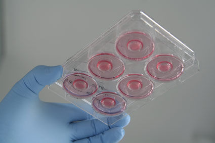
 Symbolic image: Cell culture dish with cultured skin cell cultures © BIOPRO Baden-Württemberg GmbH I Bächtle
Methods similar to those used for the regeneration of cartilage can in principle also be used for the regeneration of other tissue, including skin, bones and fatty tissue. However, the generation of artificial skin, bone and fatty tissue is associated with the problem of vascularisation and the supply of the tissue with nutrients. Cartilage tissue does not contain blood vessels, and nutrients and metabolic by-products are exchanged by passive diffusion. Therefore, joint movement, i.e. physical activity, is essential for the maintenance of normal articular cartilage.
At present, bone and fatty tissue defects can only be treated using tissue-engineered material when the lesions do not exceed one or a few centimetres in size. This is because the blood vessels cannot supply nutrients and oxygen to a larger area. The co-cultivation of tissue cells with blood vessel cells is a promising approach in the effort to also treat larger fatty tissue lesions. Studies have shown that bone defects, in the face or jaw for example, can be treated more effectively with metal implants seeded with specific organic compounds or cells. A group from the University Centre of Dentistry, Oral Medicine and Maxillofacial Surgery in Tübingen is currently working on this. Researchers from the Hohenstein Institute have shown that textile implants seeded with stem cells are also suitable as a matrix from which new tissue can be developed and used for treating cardiac and abdominal defects (see also: Hohenstein researchers make progress on biotolerance of textile implants and Hohenstein researchers successfully colonize a textile implant with human stem cells). These innovative methods are excellent examples of the biologisation of medical technology that is likely to be used for a much broader range of medical applications in the future.
Synthetic vessels with living inner walls
The coating of implants generally plays a key role in the regeneration of vessels and tubes in the human body, whether this concerns blood vessels, the oesophagus or the trachea. Research has shown that the biological coating of synthetic vessels and tubes makes the implants more compatible with the recipient’s body and blood. A smart method is the colonisation of the inner vessel walls with autologous, i.e. the patient’s own cells, which represent the basis for growing a replacement epithelium that is as natural as possible. Doctors and researchers from the Department of Cardiac, Thoracic and Vascular Surgery at the University Hospital of Tübingen are working with researchers from the Reutlingen-based NMI Natural and Medical Sciences Institute and other partners on the development of a special technology that enables them to anchor short nucleic acid strands (aptamers) on the inner surface of blood vessels. These aptamers serve as capture molecules that specifically recognise and retain stem cells from the blood that will later develop into epithelial cells. JOTEC GmbH, a medical device company based in Hechingen, is offering solutions for the therapy of vascular diseases using artificial blood vessels and stent grafts. JOTEC also works with researchers from the University Hospital in Tübingen (see JOTEC – artificial blood vessel specialist).
Bioreactors create excellent growth conditions
Bioreactors are important tools in the field of tissue engineering. Researchers at the ZRM Centre for Regenerative Biology and Medicine, a joint facility of the University Hospital Tübingen and the School of Medicine of the University of Tübingen, are working on the development of a bioreactor for the production of regenerative cornea implants. The reactor involves the use of a specific aerosol technology, which will also be used in the future for the three-dimensional cultivation of other tissues (Innovative 3D bioreactors for higher cell and tissue quality). Researchers at the Fraunhofer IGB in Stuttgart are working on bioreactors in an attempt to produce skin and liver tissue. The work has progressed enormously and the researchers are now able to use their laboratory modules to realistically test the effect of pharmaceuticals on human tissue. These examples show that regenerative methods are not just highly suitable for direct medical applications, but also for the development of medical test systems.
The Department of Cardiac Surgery at Universität Heidelberg is developing bioreactors with the visionary goal of being able to grow an entire heart (see A vision of the future: whole-heart tissue engineering) from a patient’s own heart cells. Researchers and doctors from the University Hospital of Tübingen and from the Stuttgart-based IGB are working jointly on the possibility of regenerating cardiac valves and heart muscle tissue. One of the IGB's senior researchers also teaches and carries out research at Tübingen University, thereby bringing together basic and applied research and support the effective development of vital implants (Bridging the gap between academia and industry).
A special case – the regeneration of nerves
Researchers have long believed that the central nervous system is – at least for the most part – incapable of regeneration. However, state-of-the-art neurological research has shown that human nerve function has the potential to regenerate after injury to the nervous system, particularly when stimulated by external factors. It has been shown that siRNA (small interfering RNA) is able to silence proteins that prevent the growth of neurons. Researchers at the Reutlingen-based NMI are working on methods that use siRNAs to prevent fibroses and the formation of scars.
The NMI and its partners are working on the development of nerve-guidance channels that enable peripheral nerves to grow in the desired direction and find their final destination (see Bioresorbable guidance channels – new approaches to nerve regeneration). Special cases, for example the regeneration of a network of nerves following prostate surgery, require a special matrix on which chondrocytes can grow. A team of researchers from the Department of Urology at the University Hospital of Tübingen and the NMI in Reutlingen is working on the development of hydrogels that are suitable for use as stabilising 3D matrix for innovative cell therapeutics.
A team from the Department of Orthopaedics, Trauma Surgery and Paraplegiology at Heidelberg University Hospital is working on finding out how stem cells can be used to reconnect severed nerves (Heilen Stammzellen die Querschnittslähmung?; article only available in German). Neural stem and precursor cells are also the basis for the regeneration of the gastrointestinal tract in the enteric nervous system. Researchers at the Tübingen-based ZRM are investigating and developing therapeutic options for the treatment of numerous diseases. Their major focus is the treatment of Hirschsprung disease, a disease of the gut caused by the failure of the neural crest cells. (see When the “second brain” fails – therapeutic options from the field of regenerative medicine)