Vicinity Bio: Optimisation of cancer diagnostics
Comprehensive histological diagnostics through high-dimensional imaging and artificial intelligence
Microscopic examination of tissue samples is essential, particularly in tumour diagnostics. The Tübingen-based company Vicinity Bio leverages cutting-edge imaging technologies combined with machine learning to generate comprehensive datasets of individual cells from tissue sections. This approach not only helps identify more targeted therapies but also enhances our understanding of cellular functions and processes within tissues and tumours.
The motto ‘faster, higher, further’ applies not only to sports but also to the analysis of medical samples. Researchers are constantly refining each step of the process to extract and analyse maximum data from minimal amounts of blood or tissue, in the shortest possible time. Fast, accurate and comprehensive diagnoses are especially critical when treating tumours and serious infections. However, this all requires close collaboration across various disciplines.
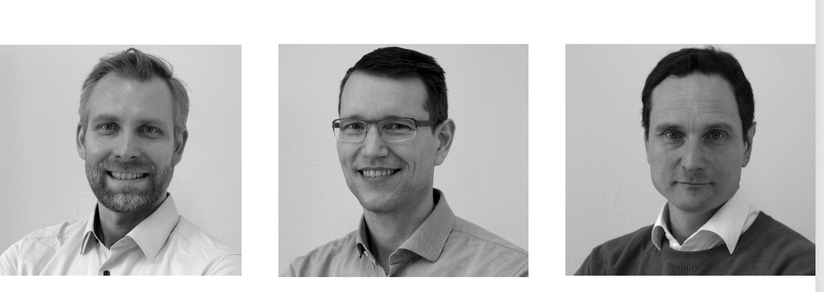
 PD Dr. Kilian Wistuba-Hamprecht, Prof. Dr. Dr. Christian Schürch and Prof. Dr. Manfred Claassen (from left to right) are the scientific founders of Vicinity Bio, a Tübingen-based company. Vicinity Bio leverages cutting-edge imaging technologies combined with artificial intelligence to generate high-dimensional images and detailed single-cell datasets from tissue samples. © Vicinity Bio
Vicinity Bio, a company founded in Tübingen in November 2024, now offers a unique service in the field of histology, i.e. the microscopic examination of tissue samples. "We offer a one-stop service from processing sample material to visualising a variety of biomarkers and AI-driven data analysis," says biochemist and immunologist PD Dr. Kilian Wistuba-Hamprecht, the start-up’s CEO. He founded the company with two other scientists, namely pathologist and tumour immunologist Prof. Dr. Dr. Christian Schürch (CMO) and clinical bioinformatician and biochemist Prof. Dr. Manfred Claassen (CTO). "Our range of skills in different fields offers extraordinary potential, which we want to utilise beyond scientific cooperation and make it available to a broad audience. We focus on both research and patient care."
Comprehensive immunohistochemical staining of tissue sections
Biological tissue is composed of a network of similar or different cells that work together to perform specific functions, and the extracellular matrix that surrounds the cells. To preserve the tissue structure as close to its natural state as possible, samples must be either shock-frozen after removal or fixed with formaldehyde and embedded in paraffin. For histological analysis, the tissue is then sliced into extremely thin sections (less than 5 µm), mounted on microscope slides, and stained to ensure that all structures are distinctly visible under the microscope
In addition to traditional chemical dyes, which are primarily used to visualise various cell components such as nuclei, mitochondria or collagen fibres, modern histology increasingly relies on antibody-mediated staining techniques. These antibodies bind specifically to target molecules, known as antigens, which are usually proteins. When antibodies are coupled with fluorescent dyes (fluorochromes), they emit signals that can be detected using a fluorescence microscope. This method is highly sensitive and specific, allowing even small amounts of antigen to be identified and quantified. It is particularly effective for analysing molecular biomarkers - molecules that reflect biological processes within cells and are therefore valuable indicators of cellular properties and functions.
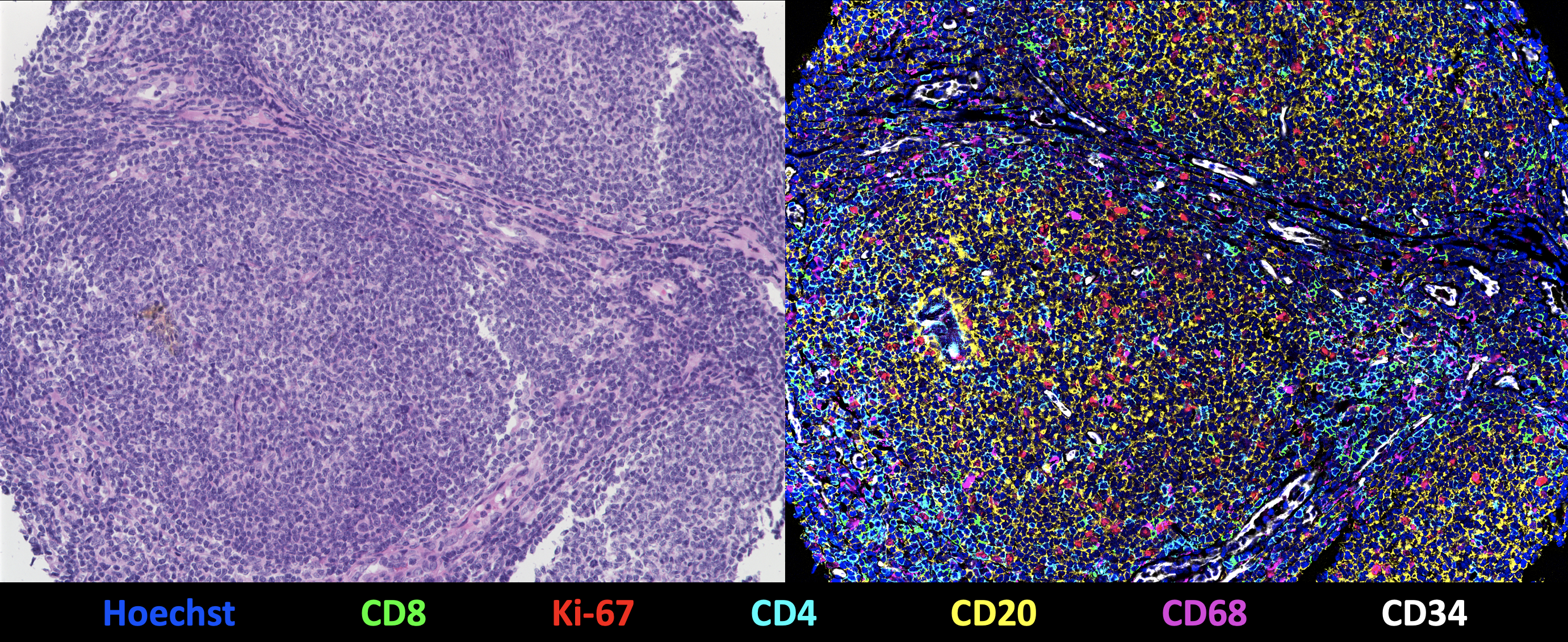
 Visualisation of the tissue nucleus of a follicular lymphoma using a CODEX multi-cycle reaction with 55 protein and 2 nuclear markers. Left: Traditional haematoxylin & eosin staining; right: 7-plex overlay image with Hoechst (cell nuclei, blue), CD8 (cytotoxic T cells, green), Ki-67 (proliferation marker, red), CD4 (T helper cells, cyan), CD20 (B cells / lymphoma cells, yellow), CD68 (macrophages, magenta), CD34 (vessels, white). The other 49 markers are not visible. © Vicinity Bio
Vicinity Bio leverages cutting-edge imaging techniques, such as high-multiplex tissue imaging (HMTI1)), to visualise a vast array of biomarkers in tissue samples. "We can detect up to 200 markers on a single tissue section, generating a high-dimensional image," explains Dr. Wistuba-Hamprecht, whose research team is now based at Heidelberg University and the German Cancer Research Center after many years at Tübingen’s Faculty of Medicine. "Compared to traditional methods, which typically capture only 6 to 7 markers, this approach offers unprecedented insights into tissue structure." This is achieved through iterative staining cycles, where up to three biomarkers are visualised at a time. After each measurement, the fluorescent dyes are removed and new biomarkers are stained in the next cycle. This automated process can be repeated 50 times or more, enabling detailed and comprehensive tissue analysis.
The scientists use platforms such as the MACSima™ system from German manufacturer Miltenyi Biotec and the PhenoCycler-Fusion (CODEX®) technology from Akoya Biosciences to analyse tissue sections. The latter employs antibodies that are not directly linked to fluorochromes but are instead temporarily labelled through a short DNA adapter sequence. This innovative technique was developed in Prof. Garry Nolan‘s laboratory at Stanford University, where both Claassen and Schürch gained extensive expertise during their years of research.
Single cell analysis using artificial intelligence
The data collected in each imaging cycle is compiled into a multi-coloured image that shows individual cells and their surrounding structures in great detail. "These colourful images are visually stunning but quite challenging to interpret," says Claassen, who is also head of the Department for Clinical Bioinformatics at the University of Tübingen’s Faculty of Medicine. His research focuses on the intersection of medicine, biology and computer science, with a particular emphasis on single-cell data for medical application. "We are developing machine learning tools capable of identifying diagnostically significant patterns within these high-dimensional images." The raw data is processed to generate a comprehensive dataset for each individual cell, which enables cell types to be identified, among other things. This approach not only detects tumour cells but also accurately identifies neighbouring cell types.
Schürch explains: "The microenvironment has a decisive influence on tumour development. Our detailed information about neighbouring cells improves diagnostics enormously and enables targeted, personalised therapy." He also sees great opportunities to identify new diagnostic and predictive biomarkers, particularly for cancer immunotherapies.
Challenging development of antibody panels
Data quality is heavily dependent on the choice, combination and sequence of antibodies. Significant expertise is required to develop effective antibody panels based on the binding properties of antibodies and the distribution of their respective antigens. "Numerous adjustments can be made, and this is where our expertise lies," Wistuba-Hamprecht points out. "We have successfully established several panels that can be adapted for different medical and scientific inquiries." Since most clients have little or no experience with this innovative technology, maintaining a close dialogue and providing extensive support throughout the process is essential. Detailed reports are typically generated within 2 to 4 weeks after receipt of a sample.
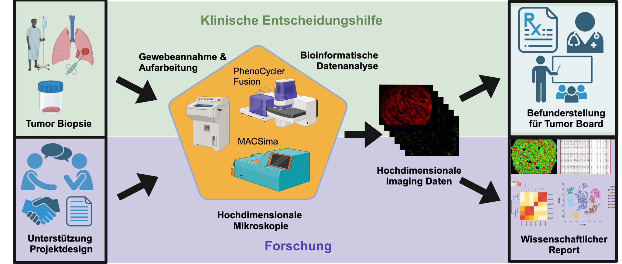
 At Vicinity Bio, all processes are consolidated under one roof, encompassing everything from the processing of tissue samples and visualisation of diverse markers to AI-driven data analysis and the generation of medical findings or scientific reports. The company also provides project design support, ensuring a comprehensive approach to research and diagnostics.
© Vicinity Bio
Award-winning concept
Dr. Aaron Mayer, co-founder of the U.S.-based company Enable Medicine, which specialises in high-dimensional image analysis, played a pivotal role in the foundation of Vicinity Bio. Moreover, the scientists at Vicinity Bio continue to work closely with the manufacturers of the imaging platforms to ensure the best outcomes.
In July 2024, the founders' innovative concept earned them first prize in the annual Science2Start ideas competition organised by BioRegio STERN.2) This recognition not only provided further support for their entrepreneurial journey but also facilitated access to a robust regional start-up network.
Vicinity Bio is committed to remaining an owner-managed business. "Our focus is on organic growth, with the goal of strengthening Cyber Valley in Tübingen over the long term," says Claassen. "We are actively seeking additional investors to support us in this mission."